Mar 2, 2024
Dr (Prof) VS Mehta adopts new technology to treat Deep Seated Brain Tumours

The brain is regarded as the most complex organ of the body. The entire functioning of the body is through the brain. Any imbalance in the same can be catastrophic for the body. In India, every year 40,000-50,000 people are diagnosed with brain tumour, of which 20 per cent are children. Some of the tumors that are detected are located in sensitive areas, where the slight complication or error could result in permanent paralysis. These are called- Deep Seated Tumors. To treat such tumors, specialized equipment, trained staff and expert team of neurosurgeons is required.

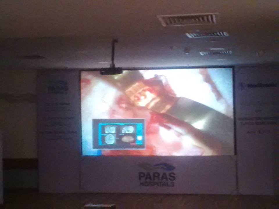
Dr (Prof) VS Mehta, Padamshree, Former HOD of AIIMS New Delhi has revolutionized Neurosurgery and medical science with a number of his achievements. His latest is the successful adoption of Image Guided Tumour Navigation Technology in the treatment of Deep Seated Tumours.
According to Dr (Prof) VS Mehta, “We have been using this technology for the last 2 years in Paras Hospitals, Gurgaon. We were the first hospital in Delhi NCR to adopt a technology that acts as the GPS for the brain. The technology is surreal as it helps the neurosurgeon to map the entire brain, pin point the affected region that has the tumour and then navigate the surgery for the best result. It is like a navigation tool for the neurosurgeons that helps us to reduce any surgical errors, there by aiding clinical outcomes.”

When Sanjiv Mishra, aged 45, as having uncontrolled headaches, he decided to see a neurologist. A brain scan detected a tumour, the size of a golf ball, deep in the brain. He and his family had apprehensions. “His surgery would have been a complicated one without the technology because of the tumour location. It was in the ventricle, a fluid space in the central part of the brain. But now deep seated tumours can be surgically removed with precision,” adds Dr (Prof) VS Mehta.
Dr Amit Srivastav, Consultant Neurosurgery, Paras Hospitals, highlights that a combination of technology support is used, “First we feed the MRI Scans of the patient in the computer. It helps map the brain. It’s like having an X ray Vision in the operating room. In addition to the anatomy, we also see the brain functioning, hence the cortical brain mapping keeps a check on the electrical impulses from the brain. This helps us monitor the sensation and the movement. The MRI puts the missing pieces of images together.”

He also adds, “To ensure that no additional pressure is being put on the brain , we use tubes through which instruments reach the tumour. The tubes are able to maneuver their way easily. The combination of everything makes a safe surgical corridor.”
Post surgery Sanjiv Mishra, feels relieved, “I can’t thank my stars enough. Going through a surgery is scary enough, but when you are safe hands you can feel confident of an easy recovery.”
Over the years Paras Institute of Neurosciences under the guidance of Dr (Prof) VS Mehta have treated hundreds of deep seated brain tumour patients. The institute has the region’s biggest neurosurgery team and is adept to handle vascular, endovascular, stereotactic and functional neurosurgery. They specialize in onco neuro surgery, skull base and spinal neurosurgery. They also have numerous successful cases of pediatric neurosurgery and peripheral nerve surgery.


